A research team from the Massachusetts Institute of Technology (MIT), Harvard University, and Massachusetts General Hospital has developed an artificial intelligence (AI) software capable of automatically analyzing white matter fiber tracts in the brainstem that were previously difficult to image clearly. This achievement opens a new window for studying the brainstem and provides a novel tool for researching neurological diseases and brain injuries. The study was published in the Proceedings of the National Academy of Sciences of the United States of America (PNAS).
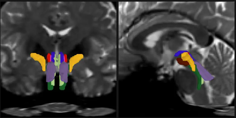
The software, named Brainstem Bundle Tool (BSBT), is based on diffusion magnetic resonance imaging (dMRI) data and uses convolutional neural networks to automatically identify and segment eight distinct white matter fiber tracts in the brainstem. Previously, due to the complex structure of the brainstem and interference from physiological motion and cerebrospinal fluid, traditional imaging techniques were unable to precisely characterize these fiber tracts.
The research team first trained the algorithm using 30 high-resolution imaging datasets from the Human Connectome Project and validated its segmentation accuracy through post-mortem anatomical results. Tests demonstrated that BSBT shows high consistency across scans of the same subject at different time points and is applicable to multiple types of imaging datasets.
In practical applications, the team used BSBT to analyze brainstem images of patients with Alzheimer’s disease, Parkinson’s disease, multiple sclerosis, and traumatic brain injury. The results showed that different diseases exhibited distinctive patterns of change in fiber tract volume and fractional anisotropy (FA) scores, which could be used for differential assessment. For example, Parkinson’s disease patients showed decreased structural integrity in specific fiber tracts, while multiple sclerosis patients exhibited simultaneous reductions in both volume and structural metrics across multiple tracts.
Moreover, in a long-term comatose traumatic brain injury patient, consecutive scans over seven months clearly showed gradual lesion shrinkage and fiber tract restoration to original positions, highlighting the tool’s potential value in assessing neural repair and prognosis.
The relevant algorithm has been publicly released for research and clinical use. The team stated that this tool not only helps deepen the understanding of the brainstem’s role in consciousness, respiration, and heartbeat, but also provides a novel imaging biomarker for early diagnosis and longitudinal monitoring of neurological diseases.
Source: Science and Technology Daily